
Introduction
Skin tag removal methods differ in how they separate or destroy a growth and in the type of wound they leave behind. At Millennium Clinic Kuala Lumpur, the choice begins with confirming that the lesion is a skin tag and assessing its size, base, location and symptoms. A method that suits a narrow stalk on the neck may not suit a broad growth close to the eye.
Snip removal, electrosurgical techniques and cryotherapy are recognised medical options, but none is universally best. Patients comparing skin tag removal methods in Kuala Lumpur should consider diagnosis, tissue preservation, healing and pigment risk alongside convenience. This article explains the practical differences without suggesting that every technique is offered by every clinic or suitable for every growth.
Key Takeaways
| Key Point | Summary |
|---|---|
| How is a method selected? | According to diagnosis, attachment, size, site and patient-specific risks. |
| Snip removal | Physically removes a suitable lesion and can preserve tissue for examination. |
| Electrical methods | Use controlled heat or electrical energy; terminology and equipment differ. |
| Cryotherapy | Freezes selected tissue, which may blister or separate later. |
| Is there a wound? | Removal creates a treated area that needs method-specific care. |
| Can a mark remain? | Yes. Pigment change or a scar is possible with any tissue-removing approach. |
Diagnosis Comes Before the Device
A wart, mole or another lesion can resemble a skin tag. Confirming identity determines whether it is reasonable to remove the growth cosmetically, leave it alone or obtain tissue for diagnosis. A changing, ulcerated or unusually pigmented growth should not be treated automatically as a routine tag. Destructive treatment can remove information that would otherwise be available to a pathologist.
The NCBI clinical reference on acrochordons discusses assessment and treatment of these benign growths. The practical implication is that a device name cannot replace clinical judgement. Patients may arrive requesting laser or freezing, yet the clinician may recommend examination, observation or an excision that preserves a specimen. Ask what finding led to that recommendation.
What Affects Method Selection?
A narrow stalk may be easier to separate than a broad attachment. Large lesions can have a different bleeding risk from tiny ones. Location matters because the eyelid, groin, underarm and neck have different nearby structures, movement and friction. The number of lesions also affects comfort, wound-care burden and whether treatment is best completed in one visit or several.
Medical history adds another layer. A tendency to form raised scars, previous pigment changes, diabetes, immune suppression or medicines that influence bleeding may alter the plan. Anaesthetic allergy and earlier removal experiences should be discussed. A doctor should explain how these factors affect the proposed method instead of offering the same treatment solely because it is the clinic’s usual equipment.
Snip Removal
Snip removal physically separates a suitable skin tag using a sterile cutting instrument. The clinician may use local anaesthetic according to the lesion and site, then control bleeding. The tag is removed during the appointment, but the small wound still needs care. Immediate absence of the growth should not be confused with immediate completion of healing.
One potential advantage is preservation of tissue when laboratory examination is appropriate. However, not every tag needs histology, and a larger or broad-based lesion may need a different surgical approach. Patients should ask whether a specimen will be sent and how results will be communicated. Snip removal is a medical technique, not permission to use household scissors on a lesion at home.
Electrosurgery and Electrocautery
Electrical treatment terms are often used loosely in advertisements. Electrosurgery uses electrical energy in tissue, while true electrocautery uses a heated instrument. Different devices and techniques can cut, coagulate or destroy tissue. The clinician should explain the approach being used rather than relying on the general word “cautery”. Settings, contact and depth affect the treated area.
An electrical method may be useful for selected small lesions or for controlling bleeding, but thermal injury can leave a crust and a temporary or persistent pigment change. Tissue destroyed by heat may not provide the same specimen as a clean excision. If you have an implanted electrical device, mention it before treatment so the clinician can assess the equipment and necessary precautions.
Cryotherapy
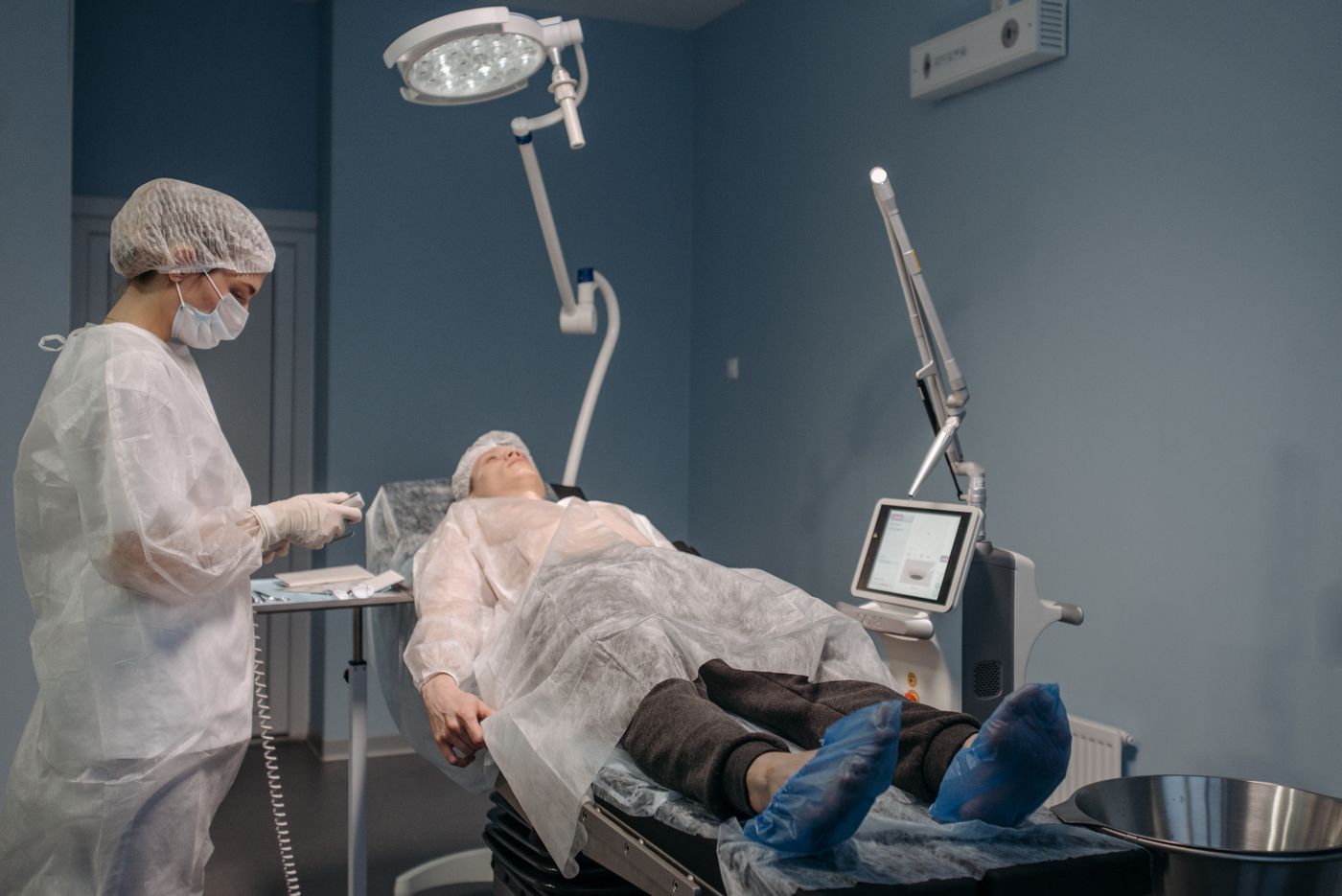
Cryotherapy applies controlled freezing to selected tissue. The treated growth may not disappear immediately; swelling, a blister or a crust can develop before separation. The extent of injury depends on factors such as freeze duration, lesion characteristics and the technique used. A review may be required if treatment is incomplete or the healing pattern is unexpected.
Freezing may cause a lighter or darker mark, particularly in pigment-prone skin. It is not automatically gentler simply because no cutting instrument is used. Nearby structures must be protected, and sensitive sites require particular judgement. Commercial home freezing kits are not equivalent to a diagnosis-based medical procedure and should not be used on an unidentified lesion or close to the eye.
A Practical Comparison
| Dimension | Snip removal | Electrical method | Cryotherapy |
|---|---|---|---|
| Mechanism | Physical separation | Controlled cutting, coagulation or destruction using the selected device | Controlled freezing |
| When the growth is removed | Usually during the procedure | Depends on technique | May separate after treatment |
| Tissue for histology | Can often be preserved | May be limited if destroyed | Usually not preserved by freezing alone |
| Early recovery | Small wound and possible bleeding | Crust, tenderness or redness | Swelling, blister or crust |
| Main trade-off | Bleeding and wound care | Thermal injury and pigment change | Delayed separation and pigment change |
The AAD lists snipping, electrical destruction and freezing as clinician-performed options. The comparison does not rank them by a single score. An appropriate choice depends on the individual lesion, and a combination of techniques may sometimes be used. Patients should be told what the planned procedure will involve and what recovery is expected for the actual site.
Where Laser Fits
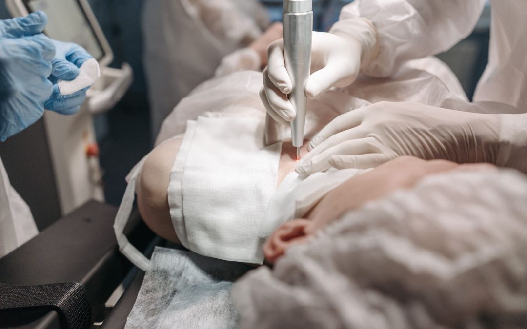
Laser treatment uses light energy to affect tissue, with the effect depending on the device and technique. It is another method offered in some practices, but “laser” covers different technologies. It should not be assumed to be scar-free, painless or suitable for every lesion. An uncertain growth still needs appropriate diagnostic assessment before destruction, regardless of how precise the equipment is described as being.
Millennium Clinic’s skin tag service page currently describes electrical and laser approaches. Snip removal and cryotherapy are discussed here as recognised alternatives, not as a claim that both are routinely available at the clinic. During consultation, ask which options are available and why the doctor recommends one for your growth. Referral may be preferable when location or diagnosis requires specialist expertise.
Is Removal Necessary?
A confirmed skin tag that does not hurt, catch or bother you may be left alone. Cosmetic preference is a valid reason to ask about removal, but it does not turn the procedure into a medical necessity. Some patients prefer to avoid a possible pigment mark or scar when the tag is barely noticeable. Others are troubled by repeated irritation and find removal worthwhile.
The decision should reflect the actual burden. A necklace repeatedly catching a tag is different from a painless growth found incidentally. Describe how often the problem occurs, whether it bleeds with shaving and what you hope to change. The doctor can then discuss whether the expected benefit justifies the procedure and whether a less irritating clothing or grooming adjustment is sufficient.
Preparing for a Removal Appointment
Bring a medication list, note previous treatment reactions and explain any attempted home removal. Avoid picking, tying or chemically burning the lesion beforehand. If the appearance has been altered, photographs from before the attempt can be useful. A clean examination is easier to interpret than one obscured by recent irritation, but do not delay assessment of a worrying change simply to allow the skin to look normal.
Ask about local anaesthetic, the number of lesions to be treated, the expected duration and whether you need help getting home. Most small procedures have a limited practical burden, but the details vary. An eyelid procedure may have different arrangements from removal of a neck tag. Clarify costs for consultation, removal, histology and follow-up separately if they are not included together.
What Happens During Treatment?
The clinician confirms the lesion and consent, prepares the area and uses the selected method. Comfort measures should be appropriate for the site. You may feel a brief anaesthetic sting, pressure or a treatment sensation, depending on the technique. Ask the doctor to explain what to report during the procedure, especially unexpected pain or symptoms near the eye.
Afterwards, the treated area may be covered or left under particular instructions. Before leaving, understand how to manage small bleeding, whether a blister is expected and when cleansing or dressing changes should start. If several lesions are treated, make sure the instructions identify any site needing different care. A verbal assurance of “minimal downtime” is less useful than a clear wound-care plan.

Healing and Pigment Considerations
Recovery depends on the wound produced, not just the size of the original tag. A shallow treated area and a deeper injury can heal differently. The NHS notes that skin tag removal can cause scarring or pigment change, including in brown and black skin. Many marks improve, but a guarantee of invisible healing would be inappropriate.
For Malaysian patients, discuss previous post-inflammatory darkening and the likelihood of sun exposure. Protection from avoidable irritation is important, especially on the neck where collars and jewellery rub. Once the surface has healed, appropriate sunscreen can support pigment care. Do not apply brightening acids or retinoids to a fresh wound unless the clinician specifically directs you to do so.
Aftercare and When to Contact the Clinic
The AAD’s wound-care guidance after skin biopsy illustrates general principles of gentle cleansing and appropriate wound protection. A skin tag procedure may need different instructions, particularly after freezing or on an eyelid, so use the plan given by your doctor. Do not automatically apply alcohol, peroxide or antibiotic cream simply because you have used them for another injury.
Increasing pain, pus, spreading redness, fever or persistent bleeding require advice. Do not deliberately pop a blister or remove a crust to make the area look smoother. If a dressing repeatedly becomes wet with sweat, ask how to manage it rather than leaving a dirty dressing in place. Record the expected review date and contact route for questions about delayed healing.
Avoiding Misleading Comparisons
Advertisements may call one technique “non-invasive” even though it destroys skin and leaves a wound. Others may imply that a newer machine produces a better cosmetic result in every patient. A useful comparison asks how the method suits the specific lesion, whether tissue can be tested and what complications the clinician is prepared to manage.
The FDA’s skin-condition health fraud guidance warns that unproven lesion-removal products can cause injury. Regulatory wording differs between countries, but a home product’s promise of a quick result does not solve the diagnosis problem. Avoid choosing an option only because it is cheap, immediate or labelled natural. A clear medical assessment is part of the value of professional removal.
Frequently Asked Questions
Which skin tag removal method is best?
There is no single best method for every lesion. Size, attachment, site, diagnostic certainty and healing risk determine the most reasonable choice.
Does freezing remove the tag immediately?
It may take time for the treated tissue to separate. Ask what changes to expect and whether a review is planned if the growth remains.
Is cautery the same as laser?
No. Electrical or heated-instrument methods differ from light-based treatment, and each has its own equipment and tissue effects.
Will snip removal leave a scar?
A mark or scar is possible. The risk depends on the wound, location and healing tendency, even when a small tag is removed carefully.
Can a removed tag be tested?
A specimen may be preserved with an appropriate physical removal method. Discuss histology before a destructive procedure if there is diagnostic uncertainty.
Is anaesthetic always necessary?
The need depends on the lesion, technique and patient. Ask how discomfort will be managed rather than assuming a method is painless.
Can multiple tags be removed together?
Sometimes. The clinician considers total wound burden, comfort and aftercare; treating every lesion in one session is not always the most practical plan.
Can I return to work afterwards?
Many patients can, but visible marks, discomfort and job-related friction may matter. Site-specific advice is more useful than a universal downtime promise.
What if I take a blood thinner?
Tell the doctor and do not stop it yourself. Medication decisions should account for its medical purpose and the procedure’s actual bleeding risk.
Does removal prevent future skin tags?
Removing existing growths does not eliminate the tendency to form new ones. A new lesion should be assessed on its own merits.
Conclusion
Comparing skin tag removal methods means comparing clinical fit and recovery, not simply comparing machines. Confirming the diagnosis, considering tissue examination and choosing an appropriate technique can make the decision safer and more understandable. Observation remains reasonable when a benign tag causes little difficulty, while troublesome lesions can be assessed for removal.
Millennium Clinic Kuala Lumpur provides doctor-led assessment and personalised advice on available skin tag procedures. Your consultation should explain the proposed method, alternatives, expected healing and follow-up so that you can decide with a realistic understanding of both benefit and possible marks.
Featured image credit: Pavel Danilyuk via Pexels. General stock illustration.